


![Request for Information – Potential [Placeholder for Prize]](https://assets.science.nasa.gov/dynamicimage/assets/science/psd/solar/2023/09/s/solarsystem_0.jpg?w=1024)
Radiation Biophysics Lab
What We Do
The Radiation Biophysics Lab uses computational and experimental methods to understand how ionizing radiation impacts health at the level ranging from single molecules to whole organisms. We analyze the individual sensitivity to ionizing radiation in humans and model organisms and the genomic associations with radiation resilience. In addition, we develop high-throughput human 3D organ models to study the impacts of space radiation and combined exposures to spaceflight stressors.

Why We Do It
Understanding how individuals respond to ionizing radiation is crucial for human spaceflight and cancer radiotherapy. We hope to help identify and mitigate the cancer, neurovascular, immune and other health risks that astronauts will face from exposure to deep space radiation on lunar and Mars missions.

Current Research Interests
- DNA damage quantification. Over the years, our laboratory has developed a strong expertise in quantifying DNA damage using markers such as p53 binding 1 protein (53BP1), gH2AX, pATM in various preparations (adherent cells, blood, in vivo tissue). Our specialty is quantifying DNA damage in human blood cells to identify individuals with sub-optimum DNA repair and thus higher sensitivity to ionizing radiation.
- Genomic associations with sensitivity to ionizing radiation. We are conducting a large study of primary immune cells from over 700 healthy donors to investigate their responses to ionizing radiation, including simulated galactic cosmic ray components. We use semi-automated, high-throughput approaches to map the dose response and time course of DNA and cellular damage after irradiation, followed by low-coverage whole genome sequencing to examine the genomic associations with radiation sensitivity.
We have recently published an associated study on identifying the genomic associations with mouse radiosensitivity, and are currently comparing mouse and human ex vivo DNA damage responses. Ultimately, we aim to identify the key mechanisms regulating responses to ionizing radiation that could be targeted for biomarker or countermeasure development. - High-content analysis, microscopy and flow cytometry. Our laboratory relies on a strong expertise in fluorescent microscopy, including super-resolution, high-throughput and confocal microscopy integrated into custom software for unbiased quantification of images and flow cytometry data. Computational biology, modeling and image processing methods are developed in the laboratory to answer multiple biological questions.
- Human organ models to understand the effects of deep space radiation. We develop human high-throughput 3D models of the central nervous system and the blood-brain barrier to investigate the impairments caused by simulated deep space radiation, with goals for expanding them to include other organ systems and applications and possible payload adaptation. We have also worked on 3D cancer models in the past and plan to integrate them in the future for evaluating carcinogenesis caused by spaceflight-relevant stressors.
- Chronic exposures to spaceflight-relevant stressors. To achieve a more accurate representation of spaceflight conditions, we have built a low dose rate gamma irradiation setup using 57Co flood source. We use this setup to analyze cellular and tissue responses to chronic exposures to ionizing radiation combined with other spaceflight stressors such as simulated microgravity and high CO2 concentration.
- Biomarkers of ionizing radiation responses. We investigate secreted markers, including cytokines and exosomes, as markers of responses to ionizing radiation and as potential personalized markers of individual radiation sensitivity. Similarly, we are also interested in omics markers: i.e. transcriptomic signature and methylation profiles.


Lab Members
Sylvain V. Costes, Ph.D.Lead Principal Investigator. Radiosensitivity lead.
Egle Cekanaviciute, Ph.D. Principal Investigator. Neurovascular lead.
Connie Tsai Pasternak, Ph.D. Staff scientist, Blue Marble Space Institute of Science.
Sonali D. Verma, B.Sc. Research associate, Blue Marble Space Institute of Science.
Estrella Passerat de la Chapelle. Research associate, Blue Marble Space Institute of Science.
Co-mentored with NASA GeneLab:
Sonali Patel. Research associate, Blue Marble Space Institute of Science.
Anmol Seth. UC Berkeley undergraduate student.

Lab Alumni

Jonathan Oribello, Research associate, Blue Marble Space Institute of Science. 2021.
Eloise Pariset, Ph.D. NASA Postdoctoral Scholar; TRISH Postdoctoral Scholar. 2018 – 2021.
Sherina Malkani. Research associate, Blue Marble Space Institute of Science. 2018 – 2020.
Alejandra Lopez Macha. Research associate, Blue Marble Space Institute of Science. 2019 – 2020.
Margaux Petay. Research associate, Blue Marble Space Institute of Science. 2020.
Vanesa Gomez Gonzalez. Software engineer, USRA. 2018 – 2020.
Roberta Boreham. Summer intern. 2019.
F. Marcus Vincent. Summer intern. 2019.
Ivan G. Paulino Lima, Ph.D. Visiting research scientist, Blue Marble Space Institute of Science. 2018.
Shayoni Ray, Ph.D. Visiting scientist, USRA. 2017 – 2018.


Publications
- Sylvain Costes: Google Scholar
- Egle Cekanaviciute: Google Scholar
- Penninckx, S., Pariset, E., Cekanaviciute, E. and Costes, S.V. (2021). Quantification of DNA damage foci to evaluate and predict biological responses to ionizing radiation. In press, Nucleic Acids Research: Cancer.
- Nelson, C.A., Uriarte Acuna, A., Paul, A.M., Scott, R.T., Butte, A., Cekanaviciute, E., Baranzini, S.E. and Costes, S.V. (2021). Knowledge network embedding of transcriptomic data from spaceflown mice uncovers signs and symptoms associated with terrestrial diseases. Life 11(1), 42.
- Malkani, S., Chin, C.R., Cekanaviciute, E., Mortreux, M., Okinula, H., Tarbier, M., Schreurs, A.-S., Shirazi-Fard, Y., Tahimic, C.G.T., Rodriguez, D.N., Sexton, B.S., Butler, D., Verma, A., Bezdan, D., Durmaz, C., MacKay, M., Melnick, A., Meydan, C., Li, S., Garrett-Bakelman, F., Fromm, B., Afshinnekoo, E., Langhorst, B.W., Dimalanta, E.T., Cheng-Campbell, M., Blaber, E.A., Schisler, J.C., Vanderburg, C., Friedlander, M.R., McDonald, J.T., Costes, S.V., Rutkove, S., Grabham, P. Mason, C.E., and Beheshti, A. (2020). Circulating miRNA spaceflight signature reveals targets for countermeasure development. Cell Reports 33(10):108448.
- Pariset, E., Bertucci, A., Petay, M., Malkani, S., Lopez Macha, A., Paulino Lima, I. G., Gomez Gonzalez, V., Tin, A.S., Tang, J., Plante, I., Cekanaviciute, E., Vazquez, M. and Costes, S.V. (2020). DNA damage baseline predicts space radiation and radio-therapeutic resilience. Cell Reports 33(10):108434.
- Afshinnekoo, E., Scott, R.T., MacKay, M.J., Pariset, E., Cekanaviciute, E., Barker, R.J., Gilroy, S., Hassane, D., Smith, S.M., Zwart, S.R., Nelman-Gonzalez, M., Crucian, B.E., Ponomarev, S.A., Orlov,O.I., Shiba, D., Muratani, M., Yamamoto, M., Richards, S.E., Vaishampayan, P.A., Meydan, C., Foox, J., Myrrhe, J., Istasse, E., Singh, N., Venkateswaran, K., Keune, J.A., Ray, H.E., Basner, M., Miller, J., Vitaterna, M.H., Taylor, D., Wallace, D., Rubins, K., Bailey, S.M., Grabham, P., Costes, S.V., Mason, C.E. and Beheshti, A. (2020). Fundamental biological features of spaceflight: advancing the field to enable deep space exploration. Cell 183(5):1162-1184.
- Penninckx, S., Pariset, E., Uriarte Acuna, A., Lucas, S. and Costes, S.V. (2020). Considering cell proliferation to optimize detection of radiation-induced 53BP1+ foci in 15 mouse strains ex vivo. Radiation Research 195(1):47-59.
- Nikitaki, Z., Pariset, E., Sudar, D., Costes, S.V. and Georgakilas, A.G. (2020). In situ detection of complex DNA damage using microscopy: a rough road ahead. Cancers 2020 Nov 6; 12(11):3288.
- Pariset, E., Penninckx, S., Degorre Kerbaul, C., Guiet, E., Lopez Macha, A., Cekanaviciute, E., Snijders, A.M., Mao, J.-H., Paris, F. and Costes, S.V. (2020). 53BP1 repair kinetics for prediction of in vivo radiation susceptibility in 15 mouse strains. Radiation Research 194 (5) 485-489.
- Pariset, E., Malkani, S., Cekanaviciute, E. and Costes, S.V. (2020). Ionizing radiation-induced risks and countermeasures in the central nervous system. International Journal of Radiation Biology 2020 Oct 20:1-19.
- Penninckx, S., Cekanaviciute, E., Degorre, C., Guiet, E., Viger, L., Lucas, S. and Costes, S.V. (2019). Dose, LET and strain dependence of radiation-induced 53BP1+ foci in 15 mouse strains ex vivo introducing novel DNA damage metrics. Radiation Research 192(1): 1-12.
- Cekanaviciute, E., Rosi, S. and Costes, S.V. Central nervous system responses to simulated galactic cosmic rays. (2018). International Journal of Molecular Sciences 19(11): 3669.
- Ray, S., Cekanaviciute, E., Paulino Lima, I., Singers Sorenson, B., and Costes, S.V. (2018). Comparing proton and charged particle therapy using DNA damage biomarkers. International Journal of Particle Therapy 5(1): 15-24.
- Beheshti, A., Cekanaviciute, E., Smith, D.J. and Costes, S.V. (2018). Global transcriptomic analysis suggests carbon dioxide as an environmental stressor in spaceflight: a systems biology GeneLab case study. Scientific Reports 8:4191.
Contact Us
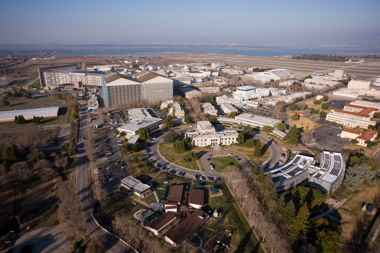
The Radiation Biophysics Lab is located at NASA Ames Research Center Collaborative Biosciences Laboratory Building N288 in Moffett Field, CA. You can reach us via email below if you have any questions about our work or ideas for collaboration.
Sylvain Costes: Sylvain.V.Costes@nasa.gov
Egle Cekanaviciute: Egle.Cekanaviciute@nasa.gov