Spaceflight Associated Neuro-Ocular Syndrome
The Challenge
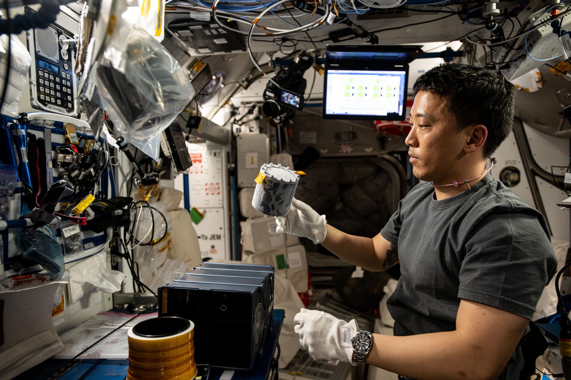
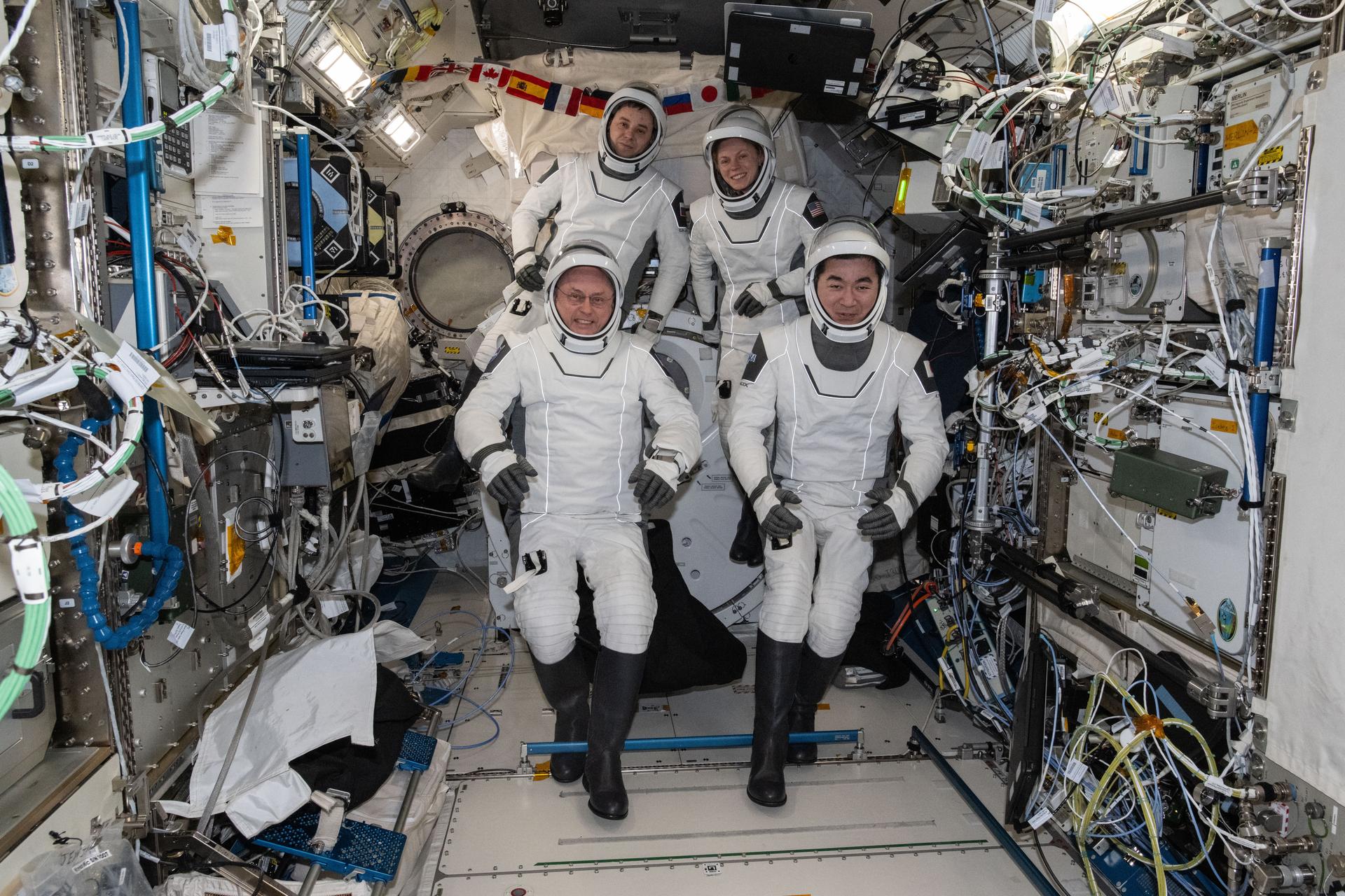
In several cases, astronauts have experienced lasting changes in their visual acuity when they returned to Earth after long-duration spaceflight missions aboard the International Space Station. Referred to as spaceflight associated neuro-ocular syndrome (SANS), no clear explanation as to why such ophthalmic changes might occur in microgravity currently exists. It has been proposed by space medicine experts that the large displacement of body fluid toward the head in microgravity, via biomechanical pathways, may be a significant contributor to some of these ocular changes by influencing the way pressure is distributed in the cranium.
The Research
The Cross-Cutting Computational Modeling Project (CCMP) has developed a suite of computational models to simulate the effects of fluid shift on the cardiovascular, central nervous, and ocular systems to understand the cause of SANS syndrome. The models rely on fundamental research and development of lumped parameter, finite element, and computational fluid dynamic numerical modeling. These models will help identify critical parameter interactions and enhance understanding of physiological responses as well as guide countermeasure development by characterizing the appropriate biomechanical response of the cardiovascular, central nervous, and visual systems in microgravity.
The Progress
The CCMP has developed models of organ systems hypothesized to contribute to SANS syndrome. Models vary in the size resolution of the structures they model, the time step used to simulate changes within those systems, and the accuracy with which the model reproduces human anatomy. These models include the whole-body model and eye model. Together, these models provide information on contributions of intracranial pressure (ICP), ocular blood flow, globe volume, intraocular pressure (IOP), and the cardiovascular system (CVS). In addition, the eye model (finite element) enhances understanding of biomechanical stress/strain and tissue remodeling that may occur during SANS. These models are used to examine key anatomical structures and physiological processes and to interpret experimental data generated to understand the cause of SANS.
Technical Data
- presentation_HSE_09182011_Final
- Myers_VIIP_abstract_draft20120510_Final
- 9th Annual SBMT – 10May2012 – compressed-FINAL
- Microgravity-induced fluid shift and ophthalmic changes
- Modeling the Effects of Spaceflight on the Posterior Eye in VIIP
- Numerical Modeling of Ocular Dysfunction in Space
- Biomechanics of the Optic Nerve Sheath in VIIP Syndrome
- Modeling and Simulation of Visual Impairment and Intracranial Pressure (VIIP) in Space
- Computational Models of the Eye and their Applications in Long Duration Space Flight
- An integrated biomechanical model for microgravity-induced visual impairment

